Physico-chemical characterization
Physico-chemical properties of new emerging nanomaterials and nanoparticulate drug delivery systems have a strong influence on their interaction with biological matter, their bioavailability and biodistribution. Therefore, the physico-chemical characteristic of nanomaterials need to be precisely defined.
We perform thorough physico-chemical characterization of the nanomaterials developed in our group and we use different techniques to determine their properties such as size, shape, surface properties, chemical modifications and more.
Morphology of nanoparticles is determined using cryo-electron microscopy (Cryo-EM) and scanning electron microscopy (SEM). Size (diameter), size distribution and polydispersity index are routinely determined by means of transmission electron microscopy (TEM) and dynamic light scattering (DLS). If required and for highly concentrated samples, small-angle X-ray scattering or ultra small-angle X-ray scattering (U-SAXS) is additionally performed to determine the size distribution of the nanoparticles.
Surface properties including zeta potential, porosity, specific surface area are characterized with methods such as electrophoretic light scattering and gas adsorption. If necessary, crystallinity of the material is checked using X-ray powder diffraction (XRPD).
Modification of nanoparticles with targeting moieties is verified by means of Fourier transform infrared spectroscopy (FTIR) and the number of fluorescently labeled conjugated ligands per nanoparticle is determined using fluorescence correlation spectroscopy (FCS). Moreover, any modification of a material with fluorescent markers is additionally confirmed with flow cytometry or confocal laser scanning microscopy (CLSM).
Depending on the system and its application, other physico-chemical techniques and methods such as circular dichroism (CD) and fluorescence spectroscopy, capillary electrophoresis (CE), specific volume calculations, critical aggregation concentration (CAC) determination are applied to further characterize new nanomaterials.
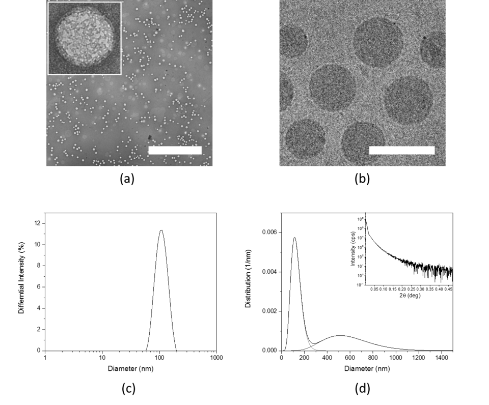
Figure: Morphology and size determination of solid-sphere PEG-b-PCL nanoparticles (PEG-PCL NP) by TEM, cryo-EM, DLS and U-SAXS. (a) TEM image of PEG-PCL NP, size bar 2500 nm; (b) cryo-EM image of PEG-PCL NP, size bar 100 nm; (c) Size distribution of PEG-PCL NP by DLS; and (d) Size distribution of highly concentrated PEG-PCL NP by U-SAXS, inlet shows measured scattering intensity as a function of 2θ angle. (a), (b), (c) 5 mg/mL and (d) 50 mg/mL.
Publications:
- Comparative safety evaluation of silica-based particles
- Functionalized Solid-Sphere PEG-b-PCL Nanoparticles to Target Brain Capillary Endothelial Cells In Vitro
- Immobilization of Enzymes on PLGA Sub-Micrometer Particles by Crosslinked Layer-by-Layer Deposition
- Formation of lipid and polymer based gold nanohybrids using a nanoreactor approach