Tiny fish under a giant camera
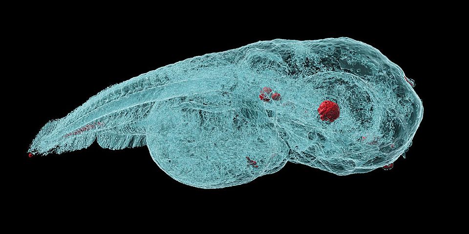
Using microtomography in phase contrast mode, researchers at the University of Basel succeeded in taking high-resolution, three-dimensional images of zebrafish embryos in which the distribution of nanoparticles (red) is visible. Also in red and easy to distinguish because of their high density are the lens of the eye and the otoliths in the inner ear – that is, tiny pieces of calcium carbonate in the vestibular system (Image: University of Basel, Jan Bolten)
Metal-based nanoparticles are a promising tool in medicine – as a contrast agent, transporter of active substances, or to thermally kill tumor cells. Up to now, it has been hardly possible to study their distribution inside an organism. Researchers at the University of Basel have used a three-dimensional imaging method to take high-resolution captures inside zebrafish embryos.
To establish the effectiveness and safety of metal-based nanoparticles for medical applications, developers must investigate the particle distribution inside a living organism and detect their accumulation. Until now, the particles have had to be labeled with radioactive or fluorescent markers. Such marker molecules, however, can influence the distribution of the nanoparticles in the organism. In addition, a high dose of particles is needed and distorts the results. Read more